Understanding the sources of error in intraoral scanning of edentulous patients to find simple and effective solutions
Introduction
Intraoral scanning of edentulous arches is a complex procedure, in which apparently minor errors can significantly compromise final accuracy.
Understanding the causes of errors is essential to improve the predictability of clinical outcomes.
👉 For a complete overview of the problem, consult the guide on intraoral scanning of edentulous arches and clinical strategies
Why is scanning edentulous people more difficult?
Unlike dentate patients, edentulous arches present:
reduced reference geometry
mobile soft tissues
limited access to the scanning field
👉 These factors make the process more sensitive to acquisition errors.
The main errors in intraoral scanning of edentulous patients
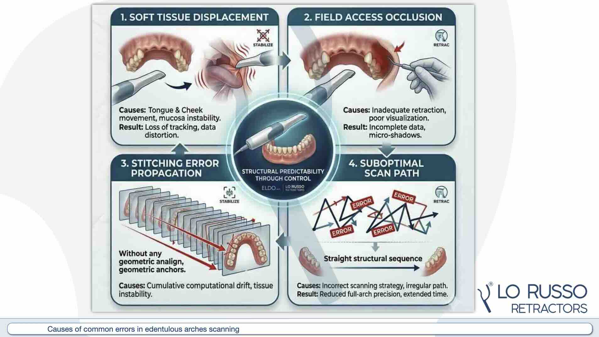
1. Instability of soft tissues
The movement of the tongue, cheeks, and mucosa is the main cause of errors.
Effect:
loss of tracking
local distortions
👉 This aspect is closely related to the clinical conditions described in the intraoral scanning of edentulous patients.
Learn how to improve tissue stability during intraoral scanning
2. Insufficient access to the scanning field
Difficult-to-reach areas lead to:
incomplete data
stitching errors
3. Stitching and tracking errors
Intraoral scanners reconstruct the image through progressive alignment.
In the absence of stability of the tissues included in the scanning field:
the system loses coherence
cumulative errors are generated
4. Suboptimal scanning sequence
The scanning strategy plays a fundamental role. An incorrect path can:
increase errors
reduce accuracy
5. Uncontrolled clinical conditions
Factors such as:
humidity
patient movement
accessibility
directly influence the quality of the scan.
How to avoid errors in scanning edentulous areas
To achieve reliable results, it is necessary to:
stabilize the soft tissues
improve access to the scanning field
use correct scanning strategies
maintain controlled clinical conditions
👉 The importance of access and stability is explored in the guide on accuracy and strategies in intraoral scanning of edentulous areas
The role of tissue stabilization
Stabilization is the key factor for:
improving tracking
reducing errors
increasing predictability
Discover tools that help you stabilize tissue and improve scan accuracy
Conclusions
Errors in intraoral scanning of edentulous areas do not depend solely on technology, but primarily on operational conditions.
👉 Checking tissue access and stability is essential for obtaining accurate and repeatable results.
To delve deeper into the topic and improve your clinical practice, consult the complete guide on intraoral scanning of edentulous arches.
Most common errors (in summary)
are due to:
- tissue instability (blocks acquisition, reduces accuracy)
- poor access (blocks acquisition, partial acquisition, artifacts)
- stitching errors
- inadequate scanning strategy
Discover the step-by-step workflow to achieve accurate results
If you want to go deeper
To fully understand how to best manage the scanning workflow of edentulous arches,
or explore these key aspects:
- Intraoral Scanning vs Conventional Impression in Edentulous: Advantages, Limitations, and Clinical Indications
- Errors in intraoral scanning of edentulous arches
- Maxillary vs Mandibular Intraoral Scanning in Edentulous: Clinical Differences and Operational Strategies
- How to stabilize tissues during intraoral scanning
- Step-by-step workflow for scanning edentulous arches
- Trueness of intraoral scanning vs conventional impression of edentulous arches
- How the quality of the scan affects prosthetic fitting
Deepen your clinical knowledge
Explore scientific articles, clinical insights, and the main challenges in intraoral scanning of edentulous arches.
Apply validated protocols
Follow step-by-step workflows to achieve predictable results in intraoral scanning and digital prosthetics.
Access courses and training
Develop advanced clinical skills with structured training paths and practical tutorials.