Intraoral Scanning in Edentulous Arches
Learn clinical protocols and soft tissue stabilization techniques to improve accuracy and workflows in edentulous intraoral scanning.
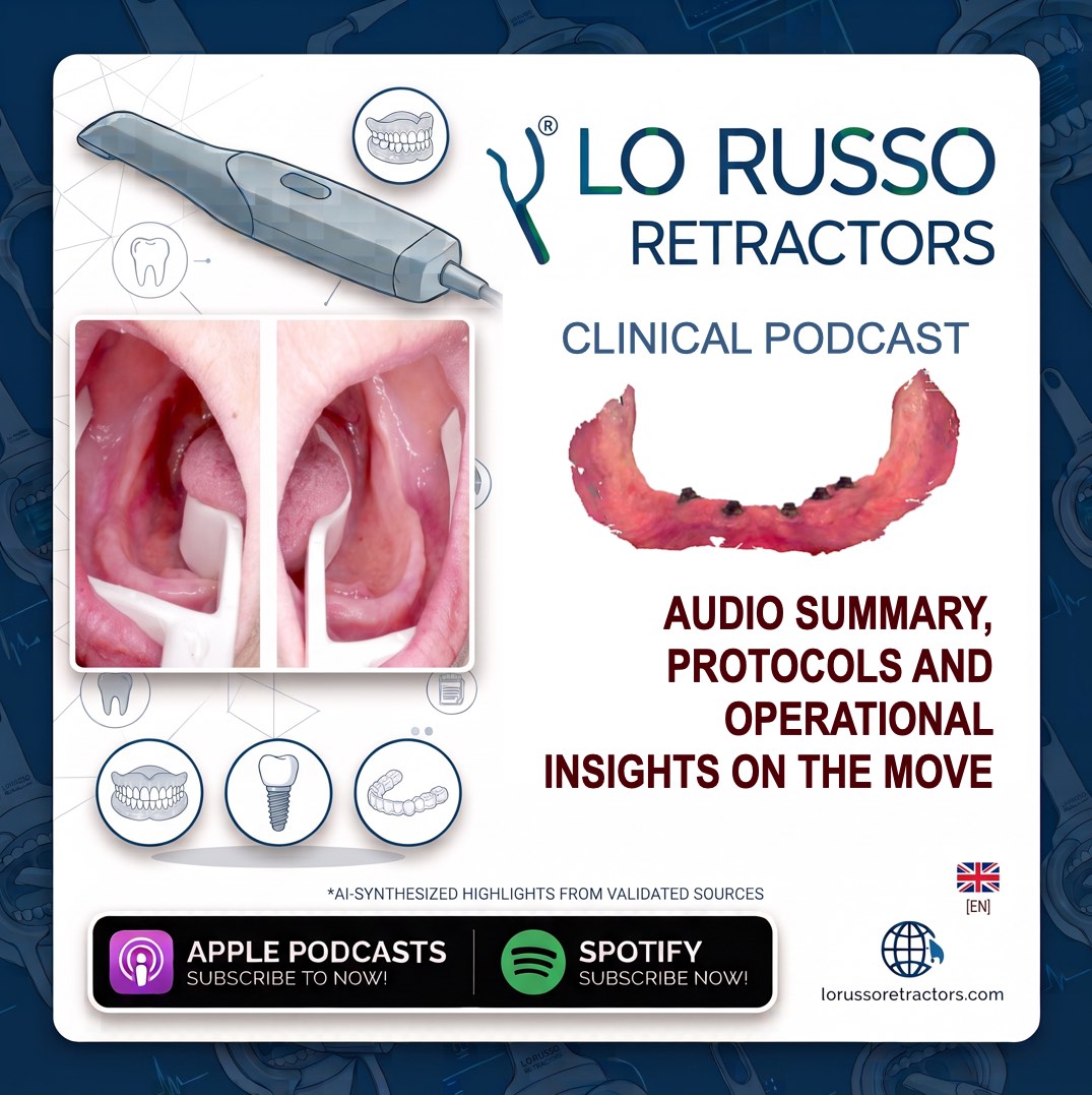
Key Episode Takeaways
Optimization of Intraoral Scanning Workflows in Edentulous Patients: Clinical Rationale and Operational Protocols of the Lo Russo Retractors System
The integration of digital workflows into reconstructive dentistry has revolutionized restorative planning, yet the optical replication of completely edentulous arches remains a significant clinical challenge. In contrast to dentate arches, which present rigid, stable anatomic structures such as enamel and fixed interproximal geometries, the edentulous oral cavity is dominated by easily displaceable, highly mobile soft tissues. Traditional impression techniques rely on physical materials that inevitably compress and deform these mucosal boundaries, often leading to unpredictable mucosal support records. Digital intraoral scanners attempt to register these surfaces in a passive state, but the lack of stationary landmarks and the constant mobility of the surrounding soft tissues introduce severe dimensional discrepancies.
For an intraoral scanner to capture a precise tridimensional digital model of an edentulous ridge, the surrounding mobile tissues must be held in a state of absolute static control, high visibility, and dry isolation. During standard scanning procedures, the tongue, labial mucosa, and buccal vestibule continuously collapse over the alveolar ridge, invading the scanner tip’s focal window. This tissue intrusion causes severe optical tracking loss, introduces motion artifacts, and generates geometric stitching errors within the reconstruction software. Clinicians frequently experience repeated scanning interruptions, prolonged chairside times, and inaccurate virtual models that ultimately compromise the fit of complete dentures, overdentures, and implant-supported rehabilitations.
Resolving these biological and mechanical challenges requires a standardized clinical protocol supported by specialized auxiliary instruments designed to stabilize the oral environment while guiding the scanner along a predictable path. Maintaining the immobility of the vestibule and the tongue is the only viable pathway to unlocking the true accuracy of intraoral scanning technology on edentulous ridges. Within this scientific framework, the patented Lo Russo Retractors® system serves as a functional, dynamic link between the intraoral scanner tip and the patient's mobile soft tissues, standardizing the acquisition process and eliminating manual retraction variables.
Dynamic Scanner Guidance and Soft Tissue Control Mechanics
The design of Lo Russo Retractors® addresses the core difficulties of edentulous scanning by establishing a stable physical relationship between the scanner tip and the alveolar ridge. Rather than acting as passive retractors, these instruments serve as a dynamic guide or track for the intraoral scanner. As the clinician moves the scanner along the ridge, the lateral arms of the instrument provide a structured path that stabilizes the sensor, maintaining the optical window in a constant, optimized relationship with the target mucosal tissues.
This guiding function is particularly effective along the vertical dimension, stabilizing the scanner relative to the Z-axis. By sliding the scanner tip directly along the engineered arms of the retractor, the operator can easily maintain the ideal focal distance and vertical height over the alveolar ridge. This control prevents sudden distance variations that disrupt software calibration, ensuring a continuous, uninterrupted data stream. The geometry of the retractors is mathematically aligned with recommended scanning strategies, establishing a workflow where the scanner path and the instrument shape work in perfect synergy.
Mandibular Soft Tissue Management and Midline Stitching Protocols
Managing soft tissues during mandibular edentulous scanning represents one of the most demanding procedures in digital dentistry. The highly active musculature of the tongue and the floor of the mouth, combined with the mobility of the buccal mucosa, presents a constant risk of ridge occlusion. The application of the mandibular retractors physically isolates the ridge from these active tissues, translating a highly dynamic biological environment into a completely static, open, and scannable field. This control exposes crucial anatomical landmarks, including the retromylohyoid fossa, the buccal vestibule, and the anterior frenula, extending the readable area almost to the tonsils.
The stabilization of these tissues is directly tied to the success of the digital image stitching process, which is the software's ability to merge individual optical captures into a cohesive, non-distorted full-arch model. To stitch the left and right halves of the mandibular arch without digital warping or duplication, the scanning software requires a highly accurate, static area of geometric overlap in the anterior midline region. The spatial orientation of the mandibular retractors is critical to capturing this reference area.
Clinicians may be inclined to hold the mandibular retractors parallel to the patient's sagittal plane, but this positioning is clinically incorrect and leads to scanning failures. To achieve proper software alignment, the retractor for the patient's left side must be held at an angle between 7 and 8 o'clock, while the retractor for the right side must be oriented between 4 and 5 o'clock. This specific angular positioning ensures that the anterior midline overlapping region is fully exposed and cleanly captured during the first half-arch scan. When the clinician switches instruments to scan the contralateral side, the software successfully identifies this stable midline overlap, using its preserved geometry to seamlessly stitch and merge the two digital files into an accurate, distortion-free mandibular model.
Standardized Clinical Workflow and Instrument Selection
Achieving predictable scanning outcomes with this system requires strict adherence to a systematic clinical protocol. Prior to patient contact, all instruments must undergo rigorous inspection to confirm structural integrity, checking for any discoloration, wear, or deformation, and must be sterilized in accordance with standard healthcare facility guidelines. The clinician must then perform a thorough clinical examination of the patient's oral cavity to ensure the absence of active mucosal lesions. Any removable dental prostheses, orthodontic appliances, or other intraoral devices that could interfere with the scanning path must be removed.
Instrument Identification and Size Calibration
Each surgical-grade kit consists of three dedicated instruments: one designed specifically for the maxilla, and two distinct instruments for the mandible, corresponding to the patient's left and right sides. To facilitate rapid chairside identification, the design of the mandibular retractors utilizes an intuitive anatomical analogy, where the curvature of the lateral arm mirrors the natural geometry of the corresponding lower arch quadrant.
Selecting the correct instrument size is a critical step in preventing mucosal compression or tissue trauma while ensuring adequate surgical field exposure. The retractors are fabricated in multiple sizes to accommodate natural anatomical variations. On the handle of each instrument, directly opposite the engraved logo, tactile and visible indicators represent the specific size. A single engraved dash identifies the minus size, which is the smallest option in the system. The size 0 instrument features a completely smooth handle surface with no raised markers. Size 1 is identified by a single raised dot, and size 2, the largest instrument in the system, is marked with two raised dots. The clinician verifies the final size selection by gently trying the instrument in the patient's mouth to ensure an easy fit and complete ridge exposure without tissue impingement.
Clinical Insertion and Stabilizing Techniques
To guarantee correct spatial orientation, the clinician must hold the retractor handle with the engraved logo facing directly toward their gaze. The insertion of the instrument's arms into the oral cavity must be performed gently using a slight rotational motion, closely replicating the clinical path used to seat a traditional impression tray. Clinicians must not bend, flex, or mechanically force the flexible arms of the instrument during insertion, as the subsequent elastic recoil could cause sudden trauma to the delicate oral mucosa.
Once positioned inside the mouth, the instrument must be seated securely in its active working position. For the maxillary arch, both lateral arms of the upper retractor must rest comfortably within the buccal vestibule. For the mandibular arch, the protocol dictates that the anterior arm is positioned laterally to the tongue to stabilize it, while the lateral arm sits in the buccal vestibule. Throughout the entire scanning sequence, the clinician must maintain a firm, steady grip on the retractor handle. The instrument must never be left loose or unsupported in the patient's mouth, as unexpected movements could cause soft tissue injury or scanner misalignment.
Material Biocompatibility and Procedural Clinical Benefits
The clinical implementation of this technopolymer retractor system offers substantial advantages across prosthetic, surgical, and restorative workflows. By establishing complete, non-yielding soft tissue retraction, the system transforms the typically unstable edentulous ridge into a static digital field. This exposure allows a single operator to perform complex full-arch scans quickly and independently, without requiring secondary assistant retraction. Furthermore, the open field provides clear visualization of crucial anatomical landmarks—such as the hamular notches and maxillary frenula—which are essential for the accurate design of complete removable dentures and implant surgical guides.
The instruments are manufactured from a medical-grade, high-performance technopolymer, selected for its high strength, biocompatibility, and thermal resistance. This advanced polymer is engineered to withstand repeated steam sterilization cycles. Standard laboratory testing and clinical monitoring confirm that these retractors can undergo at least 1000 sterilization cycles in a steam autoclave without experiencing structural degradation, dimensional changes, or loss of mechanical elasticity. This level of durability ensures high clinical safety, exceptional hygiene standards, and long-term cost-effectiveness for the dental practice. While originally developed for complex edentulous scanning, the system's clinical utility extends to partially edentulous arches, implant dentistry, fixed prosthodontics, restorative procedures, and orthodontics, particularly when treating patients with limited compliance or severe muscular hyperactivity of the tongue and cheeks.
Clinical Support and Advanced Protocols
To further optimize your digital restorative workflow, we can provide a detailed clinical protocol on managing patients with an exaggerated gag reflex using these retractors, or analyze specific scanning software settings to ensure optimal CAD/CAM integration of edentulous files.