Edentulous Intraoral Scanning Precision
Biomechanical soft tissue control and mucostatic accuracy analysis for full-arch edentulous intraoral digital scanning.
Key Episode Takeaways
Digital Intraoral Scanning of Edentulous Arches: Mastering Soft Tissue Biomechanics and Mucostatic Accuracy
The transition from traditional physical impression-taking to completely digital workflows represents one of the most significant paradigm shifts in modern prosthetic dentistry. Clinicians can now capture highly detailed three-dimensional intraoral data, bypassing the uncomfortable, time-consuming, and technique-sensitive steps associated with physical elastomer materials. In both complete removable prosthodontics and implant-supported restorations, direct optical scanning of the edentulous arch holds the promise of a fully digital sequence. This modern approach not only enhances the overall patient experience by eliminating the unpleasant gag reflex often triggered by conventional impression trays, but it also avoids the systematic dimensional changes inherent to elastic materials and the linear expansion of dental stone casts. Moreover, by digitizing the clinical workflow, dental practices can streamline laboratory communication and completely eliminate the need for physical model storage.
Despite these clear benefits, clinicians have historically expressed deep skepticism regarding the metrological trueness and predictability of edentulous digital impressions. This clinical hesitation is frequently rooted in a widespread misconception that the edentulous alveolar ridge lacks the necessary surface characteristics or geometric landmarks to guide the scanner's optical tracking sensor. Based on this false premise, some practitioners have turned to empirical and scientifically unproven techniques, such as placing auxiliary markers made of composite resin or glass ionomer cements directly onto the mucosa to help the software align the captured images. In reality, completely edentulous jaws possess stable macroscopic three-dimensional geometric landmarks, including residual alveolar concavities, convexities, and highly detailed surface textures across the hard and soft palate, which modern high-speed intraoral optical sensors are fully capable of detecting and tracking.
The true challenge of edentulous intraoral scanning is not a technological limitation of the optical hardware, nor is it an absence of stable anatomical landmarks. The actual barrier to clinical trueness is the dynamic biomechanical instability of perioral and sublingual mobile tissues. Highly active muscle structures, including the lips, cheeks, and tongue, undergo continuous, involuntary physiological and functional movement during the scanning process. In an edentulous mouth, the progressive resorption of the alveolar ridge leaves the adjacent mobile mucosa unsupported, allowing these soft tissues to collapse directly onto the residual crest. As the scanner tip moves across the arch, this dynamic tissue displacement constantly alters the oral topography in real-time, obstructing the optical pathway and introducing hidden, cumulative registration errors. Establishing physical control and absolute stability over the oral environment is therefore the essential prerequisite to unlocking the true metrological precision of digital impression-taking.
The Metrological Reality of Alveolar Ridge Topography and the Failure of Artificial Landmarks
To understand why traditional, empirical scanning modifications fail, one must examine the metrological principles of modern intraoral scanners. These optical devices project structured light patterns onto oral surfaces to calculate three-dimensional coordinates, compiling thousands of individual images into a single coordinate system. The human palate and alveolar ridges are far from featureless; their complex geometry and distinct mucosal textures provide more than enough landmarks for the software's alignment algorithms to register the surface with high accuracy. This capability is clearly demonstrated when analyzing high-resolution scans of the hard and soft palate under color-contrast visualization, which reveals a rich, highly detailed topographic map that the optical sensor can easily track without any physical assistance.
Consequently, placing artificial composite or cement buttons on the mucosa is not only scientifically unproven but is also an unnecessary procedural complication that extends chairside time without providing any metrological benefit. These artificial markings do not increase the trueness or precision of the captured mesh. Instead, they introduce plaque-retentive surfaces, complicate mucosal hygiene, and fail to address the core clinical challenge of tissue mobility. The scanner's tracking algorithms do not struggle to register the static alveolar ridge; rather, they are confused by the movement of adjacent tissues. Therefore, clinical success depends entirely on maintaining the natural, passive anatomy of the edentulous ridge perfectly exposed and static throughout the entire scanning cycle, allowing the scanner to operate at its maximum engineered speed and efficiency.
The Biomechanics of Dynamic Soft Tissue Collapse and Latent Stitching Artifacts
The dynamic behavior of perioral and sublingual mobile tissues represents the primary source of error and clinical unpredictability in edentulous intraoral scanning. The lips, cheeks, and tongue are active, highly vascularized muscle structures that continuously alter their shape and position. When the teeth are lost, the surrounding soft tissues lose their natural biomechanical boundaries, allowing the vestibular mucosa and the sublingual fold to slide and collapse over the residual alveolar crest. As the clinician guides the scanner tip along the arch, the elastic pressure of the lips and cheeks actively displaces the vestibular fornix, while the tongue moves involuntarily in response to the physical presence of the device. This continuous tissue displacement creates a changing, unstable surface that the scanner is forced to record in real-time.
As the intraoral scanner captures and stitches together thousands of individual three-dimensional frames, the constant movement of these adjacent mobile tissues confuses the software's frame-alignment algorithms. This biological interference generates hidden stitching errors and geometric distortions that are completely invisible on the monitor during the scanning session. Because the software attempts to match changing tissue coordinates, it creates a distorted polygon mesh that appears clean and complete on the screen but contains significant dimensional errors. These latent registration distortions only manifest later during the trial denture evaluation or the final denture delivery. The resulting prosthetic device frequently exhibits a poor marginal fit, compromised suction, or total biomechanical instability, requiring extensive chairside modifications, selective grinding, or complete remakes that erode clinical profitability.
Passive Biomechanical Control and the Lo Russo Retractors® Clinical Protocol
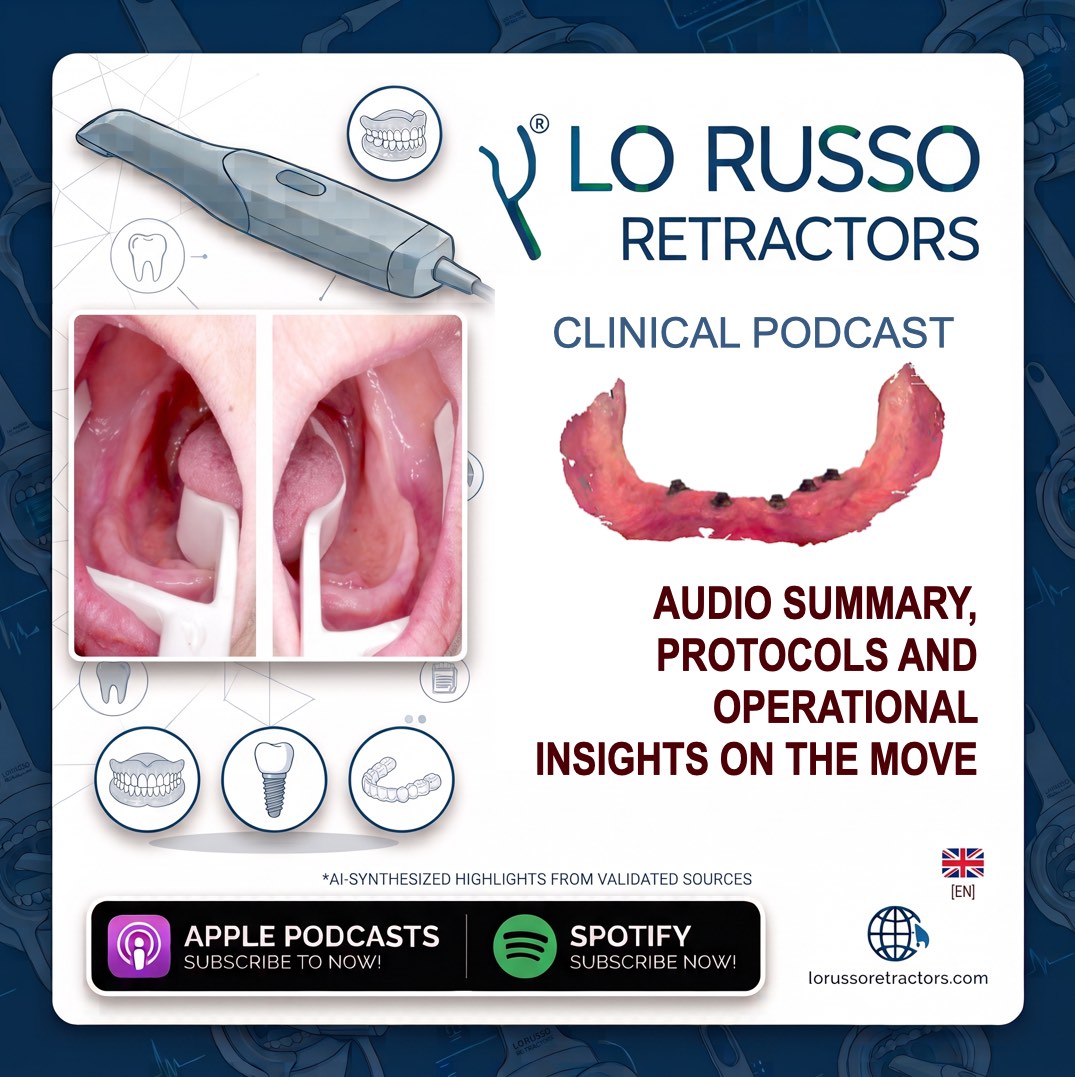
Overcoming the biological barriers of edentulous scanning requires shifting focus from software-based compensation to active, mechanical field management. The patented Lo Russo Retractors® system addresses this anatomical challenge by providing a passive biomechanical solution that matches the precise morphology of edentulous ridges. Rather than relying on aggressive, uncomfortable manual retraction, these retractors establish a gentle, continuous tension that isolates and stabilizes the entire clinical field within seconds. This system allows a single operator to perform high-speed, full-arch scans comfortably and predictably, standardizing the workflow and eliminating the clinical variability associated with manual tissue management.
The upper and lower retractors are specifically engineered to address the distinct anatomical challenges of each arch. The maxillary retractor provides highly effective passive isolation of the vestibular fornix, successfully counteracting the elastic tension of the upper lip and cheeks to keep the entire alveolar ridge exposed. This allows the optical sensor to capture critical load-bearing landmarks, including the maxillary tuberosities and the pterygomandibular raphe, which are indispensable for defining correct prosthetic boundaries and ensuring proper lip support. In the mandibular arch, the lower retractor provides comprehensive, three-dimensional control, actively containing the highly mobile tongue while deflecting the lower lip and buccal mucosa. This physical barrier prevents dynamic soft tissues from collapsing over the residual ridge and opens a clear, unobstructed optical pathway, allowing the clinician to easily guide the scanner tip to record deep posterior areas such as the retromylohyoid fossa.
The biomechanical stabilization provided by this system also validates the sequential two-step mandibular scanning protocol. This scientifically proven strategy involves scanning one hemi-arch first and then the contralateral side, joining and aligning both segments at the midline. Metrological data confirms that this segmentary approach does not compromise the geometric accuracy of the overall arch shape, provided that the adjacent mobile tissues are kept perfectly static with the retractors throughout the entire scanning cycle. By maintaining a stable, unmoving environment, the retractors prevent the software from generating stitching errors at the midline, ensuring a highly predictable and repeatable digital impression.
Metrological Trueness, True Digital Mucostasis, and Clinical Profitability
The clinical accuracy of intraoral scanning under stable biomechanical tissue control is validated by rigorous, peer-reviewed in vivo metrological trials. Comparative three-dimensional overlay analyses between digital scans obtained under stable retraction and conventional elastomeric impressions report an exceptionally small mean trueness deviation of only 30 microns for the maxilla and 20 microns for the mandible. This microscopic deviation of twenty to thirty microns does not represent an optical scanning error or a lack of digital trueness. Instead, it is the precise physical measurement of mucosal resilience and tissue compression caused by the pressure and viscosity of traditional physical impression materials during their polymerization phase.
While conventional physical impressions are inherently mucocompressive, intraoral scanning under stable retraction achieves a truly 100% mucostatic digital record, capturing the residual ridge in its natural, passive state at rest. Under the mucostatic concept, complete denture retention is achieved through surface tension generated by the intimate contact between the passive denture base and the uncompressed mucosa, rather than relying solely on a compressive peripheral seal. The high-precision, mucostatic nature of scans obtained with these retractors reduces the clinical need for complex, uncomfortable border molding procedures, allowing for shorter, highly comfortable denture flanges without compromising retention. Furthermore, three-dimensional overlay analyses demonstrate that digital meshes obtained under stable retraction show a clean, uniform extension that captures relaxed mucosa far beyond the residual ridges, completely free of any specific geometric distortion patterns.
Integrating biomechanical field control into the digital workflow optimizes both the clinical predictability and the financial efficiency of the modern dental practice. Eliminating latent digital distortions drastically reduces chairside adjustment times during denture delivery, minimizes laboratory modifications, and decreases prosthetic remake rates, resulting in a maximum return on investment. Furthermore, replacing large, uncomfortable physical impression trays and sticky elastomeric materials with a rapid, passive optical sweep under controlled retraction completely eliminates the gag reflex, providing an unmatched, elite experience for the edentulous patient. Manufactured from premium, autoclavable medical-grade materials, the retraction system complies with international ISO quality management standards and holds European CE medical device marking, ensuring maximum biosecurity and complete regulatory safety for the dental clinic.