Video Overview (2 Min)
Audio Overview
Listen to the quick summary of the clinical article
Discover Lo Russo Retractors® System
Explore the SystemVisual Mind Map
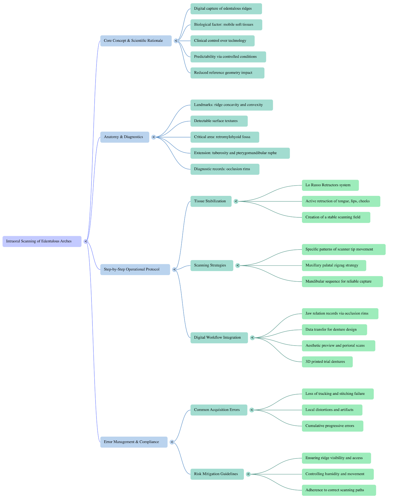
Mind map of the main concepts covered in the article - Intraoral scanning workflow and tissue control
Take Home Messages
- Optical impressions in edentulous cases are highly predictable if the clinician masters the patient's biology through validated instruments and protocols
- Control the influx of images: It is the operator's hand, stabilized by the retractors, that governs the fluidity and coherence of the images fed to the software
- Rigorous Scanning Paths: Acquisition must follow specific, structured patterns
Intraoral Scanning of Edentulous Arches: The Paradigm of Tissue Stability and Operative Field Control
In daily clinical practice, the transition toward fully digital workflows for the rehabilitation of totally edentulous patients represents one of the most fascinating, yet often feared, milestones in modern dentistry. Although the elimination of physical impressions offers undeniable advantages in terms of procedural standardization, patient comfort, and laboratory efficiency, the intraoral scanning of edentulous arches is still frequently perceived as a complex and unpredictable procedure.
However, the failure of an optical impression in full edentulism is not a problem related to the technological limits of modern scanners. It is a purely clinical and biomechanical issue strictly tied to the conditions of the operative field. It is time to analyze this challenge from a rigorously scientific perspective, debunking false myths and focusing on the true keystone of digital precision: the absolute control of the oral environment.
The False Myth: The "Lack of Reference Points"
Perhaps the most deeply rooted clinical belief is that intraoral scanners lose their orientation in edentulous arches due to the absence of teeth, which are widely considered the only valid geometric landmarks. Driven by this misconception, many clinicians resort to complex and time-consuming workarounds, such as applying drops of composite, cements, or artificial surgical markers to the mucosa.
Clinical reality and scientific data demonstrate a very different truth. Edentulous arches, as well as the tissues of the hard and soft palate, naturally present abundant macroscopic geometries (such as concavities and convexities) and detectable surface textures. Under ideal operating conditions, optical technology is perfectly capable of reading these surfaces. The use of artificial markers is not only superfluous—since their actual effectiveness has never been definitively proven—but it unnecessarily complicates the chairside workflow without solving the root cause of scanning failures.
The True Chairside Challenges: Tissue Instability and Limited Access
If the natural geometries are present and the scanner is capable of reading them, why does the data acquisition constantly interrupt? The true barriers we face every day are the unpredictability of soft tissues and the anatomical limits of the oral cavity.
The Instability of Mobile Tissues: The tongue, cheeks, lips, and non-attached mucosa are in constant movement. The software of an intraoral scanner acquires a continuous sequence of images and attempts to align them through a process called stitching. If the soft tissues change shape or position between one frame and the next, the software can no longer find the geometric matches. The inevitable result is a severe loss of tracking, the appearance of cumulative alignment errors, local distortions, and artifacts that irreparably corrupt the 3D mesh.
Limited Access and Interferences: Without adequate retraction, crucial areas of the mouth remain hidden or collapsed. The lack of direct visibility generates incomplete data acquisition and forces the clinician into continuous and erratic repositioning of the scanner.
Faced with these difficulties, the common instinct is to increase the scanning speed. But speed without control of the operative field only leads to the multiplication of errors and the frustrating need to redo the exam.
The Biomechanical Shift: Immobilize to Digitize
To transform this clinical gamble into a predictable and standardized protocol, a paradigm shift is required: moving from passive adaptation to the active management of the operative field.
Optimize your clinical workflow
Integrate Lo Russo Retractors® into your practice.
Stabilizing the soft tissues is the scientific foundation of digital dentistry in edentulous patients. By actively retracting the mobile tissues and maintaining the edentulous ridge fully and constantly exposed, parasitic micromovements of the mucosa are prevented. Only in a stable, "frozen" environment can the scanner perform its optical function fluidly and continuously, allowing for perfect image stitching.
Lo Russo Retractors®: Clinical Engineering for the Digital Workflow
To provide a definitive and validated answer to this precise clinical need, the Lo Russo Retractors® system was engineered. We are not talking about simple mouth props, but specialized medical devices designed to optimize the interface between patient anatomy and the physics of intraoral scanners.
Integrating these retractors into your clinical setup resolves acquisition problems at their root through three key actions:
Deep Anatomical Retraction: The specific design of the device completely isolates the tongue, lips, and cheeks. This provides uninterrupted visual access even in the most challenging anatomical zones, such as the retromylohyoid fossa or the pterygomandibular raphe, fully exposing the edentulous ridge.
Constant Immobilization: By keeping the soft tissues in a static and predictable position throughout the scan, sudden movements that cause loss of tracking and progressive alignment errors are entirely neutralized.
Ergonomic and Focal Support for the Scanner: The retractors act as a guiding rail on which the clinician can safely rest the scanner tip. This biomechanical feature is fundamental for maintaining an optimal focal distance from the mucosa and for standardizing the operator's movements, making them fluid and repeatable.
Clinical Accuracy: Mucostatic vs. Mucocompressive
Once the oral environment is mastered, the clinical results are measurable and highly impressive. Scientific literature has demonstrated that, by utilizing a correct retraction and scanning protocol, the optical impression of edentulous arches reaches outstanding levels of trueness.
In vivo clinical studies highlight that the mean 3D dimensional difference between an intraoral scan and a conventional impression is approximately 30 microns for the maxilla and 20 microns for the mandible.
It is essential for dental professionals to understand the true meaning of this data: we are not measuring a scanner "error". We are observing the natural physiological difference between a mucostatic intraoral scan (which captures the true resting anatomy of the tissues without physical contact) and a mucocompressive traditional impression (which deforms the tissues due to their resilience under the pressure exerted by the impression material). The absence of significant distortion patterns in the optical mesh confirms that digital digitization is clinically excellent and highly conservative for soft tissues.
Building a Predictable Digital Ecosystem
The biomechanical control of the oral cavity via Lo Russo Retractors represents the first, indispensable pillar for the transition to complete digital workflows. However, mastering the technique also requires the adoption of rigorous scanning strategies. Because it is the operator's hand that governs the influx of images to the software, following specific pathways (such as the zigzag protocol for the palate or the two-step sequence for the mandible) avoids computational overload and maximizes precision.
Scanning edentulous arches is no longer a clinical limitation, but a solid and predictable reality. Choosing clinically validated instruments means dominating the patient's biology, allowing digital technology to express its full, flawless potential.
Conceptual Summary & Workflow Steps
1. Active Tissue Control
2. Mucostatic Impression Precision
Access Clinical Masterclass
Watch complete clinical procedures, high-definition video cases, and downloadable protocols.