Intraoral Scanning Edentulous Arches Accuracy Protocol
Scientific analysis of tissue stabilization, mucostatic accuracy, and clinical protocols for intraoral scanning of edentulous patients.
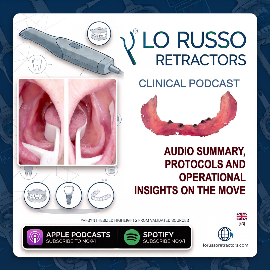
Key Episode Takeaways
Main Theme and Technological Context The transition from conventional impression-taking to direct intraoral digitization represents a major paradigm shift in modern prosthodontics, aimed at eliminating physical materials and models to improve clinical efficiency. Direct Digitization: The process of capturing oral anatomy using optical technology to establish a completely digital environment, bypassing physical casts and reducing procedural variables. The Myth of Missing Geometries: A common misconception that completely edentulous arches lack sufficient geometric landmarks to allow intraoral scanners to reconstruct a reliable three-dimensional model. Macroscopic Natural Geometries: Edentulous ridges naturally feature abundant macroscopic shapes, such as distinct curves, concavities, convexities, undercuts, and scar tissue, which provide stable reference points for the optical scanner. Microscopic Surface Texture: The oral mucosa possess a unique microscopic surface texture and fine roughness that modern high-definition scanners can detect and utilize for progressive frame-to-frame image alignment. Redundancy of Artificial Markers: The clinical application of artificial markers—such as composite resin dots, flowable materials, or metallic spheres—is unnecessary, complicates the chairside workflow, and fails to address the root biomechanical issues of soft tissue movement. Biological Implications and Clinical Criticalities Successful edentulous scanning depends on understanding the interaction between optical scanning technology and the dynamic, mobile nature of the oral cavity. Mobile Soft Tissue Instability: The constant and uncontrolled movement of the tongue, cheeks, lips, and unattached alveolar mucosa continuously alters the shape of the scanning field during acquisition. Compromised Optical Stitching: Because intraoral scanners reconstruct models by progressively overlapping sequential images, dynamic movement of tissues within the frame prevents the stitching software from establishing stable geometric matches, leading to a systematic loss of tracking. Restricted Field Access: Without physical control, adjacent mobile tissues collapse over the residual ridge, concealing deep posterior structures such as the retromylohyoid fossa, vestibular fornices, and maxillary tuberosities from the scanner's lens. The Illusion of Scanning Speed: Attempting to scan rapidly to outrun tissue movement is a clinical error that overloads the software's processing capacity, multiplies stitching errors, and introduces invisible dimensional distortions. Salivary and Light Interference: Uncontrolled saliva accumulation alters the optical refraction on the mucosa, causing scanning voids, artifacts, and inaccurate rendering of the tissue surfaces. Solutions and Operational Protocols Overcoming clinical obstacles requires a combination of structured scanning strategies and active biomechanical control of the oral tissues. The Lo Russo Retractors® system acts as an active tissue stabilization device, isolating the edentulous ridge, ensuring access to posterior structures, and serving as a stable physical track to guide the scanner tip at an optimal focal distance. Maxillary Scanning Strategy Crestal Baseline Construction: Optical capture begins at the center of the residual alveolar ridge—the most stable zone—and proceeds continuously from one maxillary tuberosity to the other, keeping the ridge centered in the scanner's viewfinder. Palatal Vault Acquisition: The scanner is returned to the midline, and the hard palate is captured using parallel, overlapping brush-like strokes, maintaining fluid movement without pausing to inspect the monitor. Buccal Aspect Recording: The vestibular and buccal sides of the maxillary crest are captured in two independent phases to simplify tissue management and prevent lip interference. Mandibular Two-Step Sequential Protocol First Hemi-Arch Lingual Progression: Following tissue drying and retractor placement, acquisition starts at the retromolar pad of the chosen side and proceeds to the midline, keeping the scanner tip slightly angled toward the lingual slope of the ridge. First Hemi-Arch Buccal Return: Upon reaching the midline, the direction of movement is reversed without stopping the scanner, progressing backward to the retromolar area along the buccal slope of the ridge. Saliva Control and Relaxation Phase: The scanner is paused and removed, allowing the patient to swallow and relax, while the clinician suctions saliva from the uncaptured hemi-arch to prevent optical reflections. Second Hemi-Arch Lingual Progression: The scanner is reinserted, and acquisition restarts strictly from the previously captured midline to enable immediate automatic mesh alignment, progressing lingually toward the opposite retromolar pad. Second Hemi-Arch Buccal Return: Without stopping the software, the scanning sequence is completed by moving from the posterior retromolar pad back to the midline along the buccal aspect, sealing the final mandibular mesh. Clinical, Biological, and Procedural Advantages The integration of structured scanning paths with active biomechanical field control provides high accuracy and clinical predictability. Micrometric Trueness in Vivo: Controlled clinical trials using advanced 3D superimposition and best-fit algorithms document a mean 3D deviation of only 30 microns for the maxillary arch and 20 microns for the mandibular arch compared to conventional impressions. Mucostatic Optical Impression: Traditional elastomer impressions are mucocompressive, deforming soft tissues under physical pressure, whereas intraoral scanning operates as a purely mucostatic technique, recording tissues in their true resting state. Tissue Resilience Quantification: The minor dimensional differences of 20 to 30 microns measured between digital and conventional impressions represent the physical displacement of mucosal tissues under the compression of analog materials rather than digital inaccuracy. Superior Anatomical Extension: Three-dimensional color-map analyses demonstrate that digital models captured under active stabilization with Lo Russo Retractors® are more anatomically extended in posterior areas (pterygomandibular raphe, tuberosities, retromylohyoid fossa) without exhibiting distortion patterns. Single-Operator Efficiency: The stabilization device manages the tongue, cheeks, and lips with one hand, allowing a single clinician to operate the scanner autonomously, reducing hand fatigue, standardizing scanner movement, and maintaining a constant focal distance. Protester Fit and Workflow Predictability: Maximizing accuracy at the impression stage directly translates to a superior passive fit of CAD/CAM prostheses, minimizing chairside adjustments, eliminating physical stone casts, and reducing remakes.